Notice
[CONFERENCE] IMPLANTEUS LECTURES Perrine Chaurand
- document 1 document 2 document 3
- niveau 1 niveau 2 niveau 3
Descriptif
20 JANVIER 2021 Avignon Université.
Using medical imaging methods in environmental sciences
- Perrine CHAURAND (CEREGE, Aix en Provence):
X-ray imaging in 2D and 3D: multi-scale and correlative approach coupling X-ray fluorescence and tomography techniques. Applications in environmental science
Perrine CHAURAND, https://www.cerege.fr/en/users/perrine-chaurand
Since 2007, engineer at CEREGE- Aix Marseille University, Aix-en-Provence. In charge of the 2D&3D X-ray imaging platform MATRIX, https://www.cerege.fr/fr/matrix
Academic background: Engineer in Chemical Engineering, Dr. in Geosciences of Environment.
Expertise: Physico-chemistry characterization of heterogeneous and divided solids (waste, nanomaterials …) with a multi-scale approach. X-ray techniques (micro-analyses, mapping and 3D imaging). Speciation of trace elements (X-ray absorption spectroscopy).
X-ray imaging in 2D and 3D: multi-scale and correlative approach coupling X-ray fluorescence and tomography techniques. Applications in environmental science.
Abstract:
2D and 3D X-ray imaging at high spatial resolution (i.e. micrometric and nanometric scale) represents the heart of the MATRIX platform located at CEREGE (Aix en Provence, France). MATRIX brings together in the same laboratory high-performance X-ray tools: two X-ray tomographs, several X-ray micro-fluorescence spectrometers and an X-ray diffractometer. These tools offer the opportunity to visualize in 2 and/or 3 dimensions the structural, the mineralogical and the chemical heterogeneity of very varied samples (geological samples, materials, biological samples, soft tissues, etc.). The use of X-ray radiation avoids the delicate and time-consuming preparations of other observation methods (such as electron microscopy) and the integrity of the samples is, in many cases, preserved.
X-ray tomography is a powerful 3D imaging technique for the in-situ and non-destructive investigation of the inner structure of an object. This relatively recent technique has tremendously evolved over the past decade with much more sensitive detection systems and increased spatial resolution. Indeed, it is now possible to reach spatial resolution of tens of nanometers with synchrotron X-ray source but also with lab-based systems (called nano-tomograph). The MATRIX platform is equipped with two X-ray tomographs, a micro-tomograph and a nano-tomograph, that offer the opportunity to perform multi-scale analysis on the same sample from several tens of microns up to 50 nm.
3D micro and nano-structural observations can also be integrated in a correlative approach by coupling X-ray tomography with 2D chemical mapping (X-ray fluorescence micro-spectroscopy, micro-XRF).
This lecture will introduce these X-ray imaging techniques: theoretical principles, operating mode and systems. Some examples of applications in environmental science (with a specific focus on the analysis of soft tissues as plants, organisms, organs or cells) will be detailed to illustrate the advantage and performance of a multi-scale and correlative approach.
Dans la même collection
-
[Implanteus Lectures] Mercredi 20 mars – « Les vésicules végétales comme vecteurs de molécules phar…
[Implanteus Lectures] Mercredi 20 mars – « Les vésicules végétales comme vecteurs de molécules pharmacologiques » par Dr. Alberto DAVALOS
-
[Implanteus Lectures] 5 mars 2024 – « Communication moléculaire entre les plantes et les microbes »
[Implanteus Lectures] 5 mars 2024 – « Communication moléculaire entre les plantes et les microbes » par Dr. Elodie Vandelle, Department of Biotechnology – University of Verona (Italy)
-
[Implanteus Conférence] 18 janvier 2024
[Implanteus Conférence] 18 janvier 2024 « Embracing social factors for soil conservation », Alexander Neaman (Chili), Professeur en gestion de l’environnement et édaphologue
-
[Implanteus Lectures] 17 janvier 2024
[Implanteus Lectures] 17 janvier 2024 « L’utilisation de sous-produits agroalimentaires » par Ana Isabel Cordeiro, Instituto Politécnico de Portalegre, Portugal
-
[Implanteus Lectures] 17 octobre 2023
Micol MolinaVicente[Implanteus Lectures] 17 octobre 2023 : « Les polyphénols végétaux dans la prise en charge des maladies métaboliques : modulation de la prise alimentaire et du métabolisme énergétique. » par Vicente
-
[Implanteus Lectures] 23 mai 2023 à 14h30 : « Utilisation des abeilles solitaires pour la pollinisa…
[Implanteus Lectures] 23 mai 2023 à 14h30 : « Utilisation des abeilles solitaires pour la pollinisation et l’apivectorat » par Professeur Ljubisa Stanisavljevic
-
[Implanteus Lectures] 14 mars 2023 : « Haricots et produits contenant des haricots : des avantages …
[Implanteus Lectures] 14 mars 2023 : « Haricots et produits contenant des haricots : des avantages nutritionnels aux connaissances et comportement des consommateurs » par Dalia El Khoury
-
[Implanteus Lectures] 18 janvier 2023 : « Bien manger »
[Implanteus Lectures] 18 janvier 2023 : « Bien manger », par Christophe Lavelle
-
[Implanteus Lectures] 16 novembre 2022 "Food for health from the consumers' perspective : How to de…
[Implanteus Lectures] 16 novembre 2022 Nicola Bordenave
-
[Implanteus Lectures] 12 octobre 2021 Tannins and pigments from grape to wine D. Véronique CHEYNIER…
[Implanteus Lectures] 12 octobre 2021
-
[Implanteus Lectures] 17 novembre : « Nutrition et changement climatique, comment ont-ils influencé…
[Implanteus Lectures] 17 novembre 2022
-
[Implanteus Lectures] 19 janvier : « Conserving agrobiodiversity and ensuring the viability of High…
[Implanteus Lectures] 19 janvier 2022


Sur le même thème
-
[CONFERENCE] IMPLANTEUS LECTURES Linda Kinkel
19 mai 2021 Avignon Université Linda Kinkel (Dept. Plant Pathology, University of Minnesota): From ecological theory to practice: microbial management for healthy crops
-
CONFERENCE La boîte translucide : un éclairage sur l’intelligence artificielle de Pierre Jourlin
16 Avril 2021 A l'occasion de la parution en mars 2021 aux Éditions Universitaires d’Avignon de « La boîte translucide : un éclairage sur l’intelligence artificielle » de Pierre Jourlin, la
-
[CONFERENCE] IMPLANTEUS LECTURES Olivier Dangles
14 avril 2021 Avignon Université Olivier Dangles (Avignon Université.) : Plant secondary metabolites and their importance to humans
-
CONFÉRENCE IMPLANTEUS LECTURES Franseco Visioli
17 mars 2021 Avignon Université Francesco VISIOLI (University of Padova) : Nutritional benefits from eating plant products
-
[CONFERENCE] IMPLANTEUS LECTURES Eric Michel
20 JANVIER 2021 Avignon Université. Using medical imaging methods in environmental sciences Eric MICHEL (INRAE Avignon): NMR and MRI to unravel the fate of contaminants in the soil Eric MICHEL
-
[CONFERENCE] IMPLANTEUS LECTURES Yvan Capowiez
20 JANVIER 2021 Avignon Université. Using medical imaging methods in environmental sciences Yvan CAPOWIEZ (INRAE Avignon): How X-ray tomography and image analysis enlarge our vision of the
-
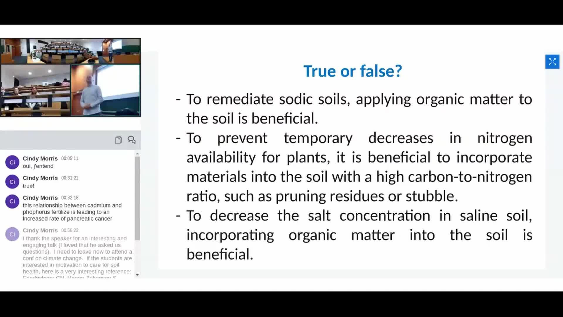
[CONFERENCE] IMPLANTEUS LECTURES Cindy Morris
17 fevrier2021 Avignon Université. Cindy Morris (INRAE Avignon)Friends or foes? Plant pathogenic microorganisms that can trigger rainfall.
-
[CONFERENCE] IMPLANTEUS LECTURES Denis COURTIER-MURIAS
20 JANVIER 2021 Avignon Université. Using medical imaging methods in environmental sciences Denis COURTIER-MURIAS (Université Gustave Eiffel): Introduction to NMR and MRI and applications
-
Communiquer par logiciel pour des personnes n'ayant pas l'usage de la parole
Cette communication a été filmée lors du colloque Angles morts du numérique, qui s’est tenu au Centre Culturel International de Cerisy du 24 septembre au 1er octobre 2020, sous la direction de
-
JPS 2019 / E-santé
Journées des Professionnels de Santé 2019 - Formation Médicale Continue des Médecins Généralistes
-
JPS 2019 / Brèves
Journées des Professionnels de Santé 2019 - Formation Médicale Continue des Médecins Généralistes
-
SEGAMED 2016 - Ouverture du congrès - Jean-Marc GAMBAUDO, Président UCA
SEGAMED 2016 Premier colloque français consacré exclusivement à la recherche et à l'évaluation des jeux sérieux appliqués à la médecine et à la santé Première Health Gaming Week appliquée à la
![[CONFERENCE] IMPLANTEUS LECTURES Linda Kinkel](https://vod.canal-u.tv/videos/media/images/universite_d_avignon_et_des_pays_de_vaucluse/.conference.implanteus.lectures.linda.kinkel_61331/vignette.jpg)
![[CONFERENCE] IMPLANTEUS LECTURES Olivier Dangles](https://vod.canal-u.tv/videos/media/images/decouvrir/.conference.implanteus.lectures.olivier.dangles_59981/vignette.jpg)
![[CONFERENCE] IMPLANTEUS LECTURES Eric Michel](https://vod.canal-u.tv/videos/media/images/universite_d_avignon_et_des_pays_de_vaucluse/.conference.implanteus.lectures.using.medical.imaging.methods.in.environmental.sciences.eric.michel_59533/vlcsnap.2021.02.16.09h54m59s341.png)
![[CONFERENCE] IMPLANTEUS LECTURES Yvan Capowiez](https://vod.canal-u.tv/videos/media/images/universite_d_avignon_et_des_pays_de_vaucluse/.conference.implanteus.lectures.using.medical.imaging.methods.in.environmental.sciences.yvan.capowiez_59511/vlcsnap.2021.02.16.09h55m15s672.png)
![[CONFERENCE] IMPLANTEUS LECTURES Cindy Morris](https://vod.canal-u.tv/videos/media/images/universite_d_avignon_et_des_pays_de_vaucluse/.conference.implanteus.lectures.cindy.morris_59595/vignette.jpg)
![[CONFERENCE] IMPLANTEUS LECTURES Denis COURTIER-MURIAS](https://vod.canal-u.tv/videos/media/images/universite_d_avignon_et_des_pays_de_vaucluse/.conference.implanteus.lectures.using.medical.imaging.methods.in.environmental.sciences.denis.courtier.murias_59531/vlcsnap.2021.02.16.09h54m22s469.png)